Chiari Malformation

When brain tissue extends out of the skull into the spinal canal this is called a Chiari malformation. These structural defects are found in both children and adults.
Depending on the type of the Chiari malformation, the tissue that sticks out could be from the cerebellum (the part of the brain that regulates muscular activity) or both the cerebellum and brain stem (which controls functions such as breathing, swallowing and heart rate).
Chiari malformation can hinder the flow of spinal fluid leading to a buildup that can block signals from the brain to other parts of the body.
Learn more about our neurosurgeon Kenneth M. Crandall, MD.
Types of Chiari Malformation
Chiari malformations are classified by their severity. Some people with the less severe form of Chiari malformation have no signs or symptoms.
Type I
The least severe form, Type I is often diagnosed late in childhood or in adulthood. In this type, parts of the cerebellum extend into the hole in the skull’s base where the spinal cord passes through, it but doesn't involve the brain stem.
The most common symptom is headache, particularly ones that occur after straining or sneezing. Other symptoms might include problems with balance and fine motor skills, numbness in hands and feet, neck pain, hoarseness, difficulty swallowing or blurred or double vision.
Type II
Type II and the other more severe forms of Chiari malformations are usually diagnosed in infancy or during pregnancy. In this form, tissue from both the cerebellum and brain stem extend into this area. This type is usually associated with spina bifida, a congenital condition where the backbone and spinal canal do not close before birth. Nerve tissue connecting two halves of the cerebellum may be absent or partially complete.
Type III
The cerebellum and the brain stem both protrude through the skull base all the way into the spinal cord area.
Type IV
In this the most severe and rare form, the cerebellum doesn’t fully develop. Parts of it are missing, and parts of the skull and spinal cord may be visible.
Diagnosing Chiari Malformations
The University of Maryland Medical Center Chiari Malformation Program team members exhaust all non-surgical treatments before moving forward with surgery. To provide a detailed diagnosis, our specialists use MRI with cerebrospinal fluid flow studies.
Surgical Treatments
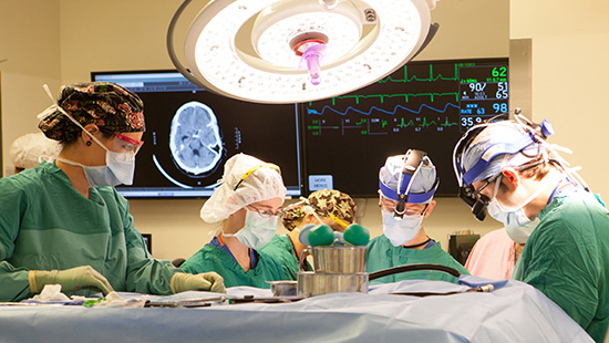
Surgery — called posterior fossa decompression — can create more space for the cerebellum and decompress the brain stem. Our neurosurgeons are experts in ensuring cerebrospinal fluid channels are open and that all tissue layers are reconstructed, resulting in more positive surgical outcomes and relief of symptoms.
